Common Surgical Procedures
Module Goals
- Learn indications for the most common surgical procedures in turtles (amputation, aural abscess drain, enucleation, and shell repair)
- Understand how to perform these surgical procedures
If you would like to listen to an audio recording of this material, please click here. (Duration: 13:44)
Introduction
While there is obviously a wide range of surgical procedures that can be performed on turtles, we will be presenting procedures that are used frequently in the Turtle Rescue Team and are important to know when dealing with wild turtles. Most of these procedures deal with the extremities, as the shell presents a barrier to more invasive intracoelomic procedures. The principles of surgery in turtles are the same as in other animals, though a few unique aspects of turtle surgery are outlined below.
- Turtles have unique anesthesia requirements – see the module on Anesthesia and Analgesia for details.
- Turtles have thin skin, and horizontal mattress sutures are the primary suture pattern of choice for skin closure.
- Suture removal is delayed in turtles, occurring at 4-6 weeks.
For advanced surgical procedures not described below, we encourage you to contact the Turtle Rescue Team for consultation or referral.
Amputation
Indications
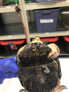
Most turtles can actually survive in the wild with only three legs. However, those requiring amputation of multiple limbs are non-releasable.
Indications for amputations include old poorly-healed fractures, open fractures with infection, tissue necrosis, muscle or ligament detachment, comminuted or multiple fractures, or a proximal fracture that cannot be stabilized and/or has potential to lead to further internal damage. If a limb has a simple fracture, is not infected, and retains sufficient neurological function, amputation is likely not necessary and the turtle should be given a chance to heal while being closely monitored for signs of infection or necrosis. The turtle might also be a good candidate for orthopedic surgery, though this would be considered a more advanced procedure.
In general, an amputation procedure in a turtle is not unlike amputations in other animals.
Procedure
- Obtain pre-surgical radiographs if possible.
- Anesthetize the turtle and give perioperative pain medication.
- 2% Lidocaine can be locally injected or topically applied at a dose of up to 10 mg/kg maximum. It is unconfirmed if lidocaine has significant analgesic effects in turtles, but it may help and will not harm them if you have it available.
- Perform dirty and clean scrubs. Dilute betadine, chlorhexidine, and alcohol/sterile saline are all appropriate.
- Make a curvilinear incision on the lateral side of the limb around the level of the middle third of the most proximal bone being removed and proximal to any infected or necrotic tissue. This will ensure enough skin and muscle will left for closure. Generally, ligation of vessels is not necessary with turtles but it can be done if profuse bleeding occurs, especially on larger patients.
- Repeat the curvilinear incision on the medial side bringing the two incisions together.
- Transect all muscle on the lateral and medial aspects until bone is exposed.
- Separate all muscle from the bone that is going to be removed until the proximal joint can be isolated.
- Incise the joint capsule and any ligament attachments to complete the amputation.
- Inspect remaining tissue and remove any necrotic or infected tissue that remains.
- Appose muscle tissue to cover the joint and use absorbable suture (2-0 to 4-0 depending on patient size) and a simple continuous or interrupted pattern to close the muscle layer.
- Close the skin using an interrupted horizontal mattress pattern and 3-0 to 5-0 absorbable or non-absorbable suture. Make some tacking bites in the muscle below to minimize dead space. Regardless of the suture type used, they will need to be removed as even absorbable sutures last a very long time in reptiles. Be sure to leave the ends long enough that they can be readily identified and removed in 4-6 weeks.
- Recover patient and apply TAO to the incision site.
- The turtle should be kept out of water for 10-14 days to minimize the chance of an infection developing in the wound. The incision can be flushed with sterile saline and TAO can be reapplied daily to aid further minimize this risk.
- Continue pain medications and antibiotics as indicated.
Aural Abscess
Indications
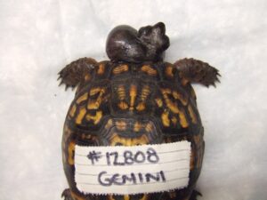
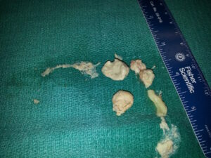
Aural abscesses are commonly seen in turtles that develop upper respiratory infections from hypovitaminosis A. In order to significantly speed up the healing process and improve a turtle’s comfort level, it is recommended that these abscesses be drained in a simple procedure. The pus in these lesions is caseous rather than liquid and may need to be scraped out with more force than expected if it is tightly packed.
Procedure
- Anesthetize the turtle and give perioperative pain medication.
- Do dirty and clean scrubs of the area with dilute betadine, chlorhexidine, or alcohol/sterile saline.
- Make a small curvilinear incision on the caudal, ventral, or caudoventral edge of the aural scale where it meets the skin.
- Use clean instruments, sterile swabs, flushing, and/or gentle pressure to remove all caseous material or as much as possible.
- Flush wound extensively and pack with TAO.
- Recover patient.
- Continue pain medications and antibiotics as indicated and continue to flush and reapply TAO until the lesion begins to heal over.
Enucleation
Indications
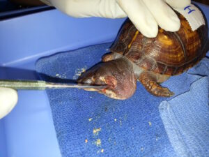
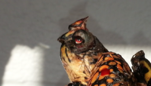
Enucleations are indicated in the case of proptosis resulting from trauma.
Procedure
- Anesthetize the turtle and give appropriate perioperative pain medication.
- Perform clean and dirty scrubs with dilute betadine and saline.
- A lidocaine retrobulbar block can be used. Typically this is at 5 mg/kg with a 10 mg/kg maximum.
- Dissect the globe from its attachments.
- If large enough to visualize, optic vessels and nerves can be ligated. Otherwise, they can be transected.
- Significant bleeding is likely so be prepared to apply adequate pressure to stop the bleeding. If hemorrhage continues, Gel-Foam can be placed into the back of the socket to aid in hemostasis.
- Be thorough in removing all excess soft tissue from the orbit.
- Trim the edges of the eyelids so that there are fresh margins that appose each other without tension.
- Close the eyelids with a horizontal mattress pattern.
- Recover patient and provide antibiotics and pain medication as indicated.
- Leave sutures in for at least 8 weeks.
Shell Repairs
Indications
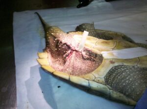
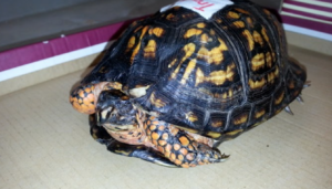
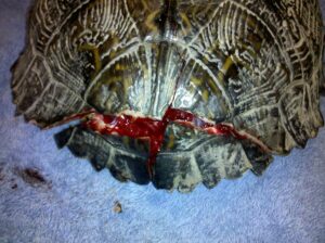
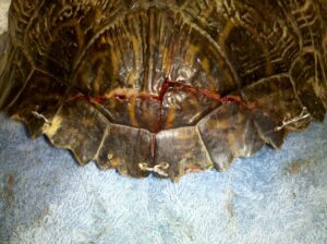
Shell repairs are the procedure most frequently performed by the Turtle Rescue Team and are indicated in cases of severe trauma, typically the result of being hit by a car. The exact procedure will vary greatly depending upon the location, severity, and number of fractures. Sometimes we have to get creative and use new materials. You are encouraged to consult the Shell Surgery and Repair chapter in the Additional Resources of this module to investigate other methods of shell repair that may align with supplies available in your practice.
These shell repair procedures are not indicated in every shell fracture case. For fractures that are well-apposed and have minimal movement, the shell can heal without hardware. Fractures that are vulnerable to being displaced by the turtle’s movements should be repaired with hardware to facilitate healing.
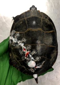
Supplies: Dremel with small drill bit, 24G wire, bra hooks, super glue, baking soda, epoxy, tongue depressor, needle drivers
Procedure
- Anesthetize the turtle and give perioperative pain medication.
- Scrub with dilute betadine or chlorhexidine and flush fractures with saline.
- Reduce fractures.
- For fractures on the edge of the carapace (drill):
- Decide where you want to place the wire ties on the edges of the carapace.
- Drill through the edge of the carapace with the dremel, placing a tongue depressor behind your drill site to stop the drill from causing tissue trauma. Make sure the holes are about 5 mm away from the edge of the carapace and the edges of fractures. Flush the holes with sterile saline.
- Feed the wire through the ventral aspect of the holes, and bring the ends together on the dorsal aspect and cross the wires as close to the carapace as possible.
- Clamp both ends of the wire with the needle drivers and rotate to twist the wires together. Do not tightly twist the fracture together at this point. Leave it a bit loose to give you room to place all the hooks before tightening anything.
- For fractures in the middle of the carapace (glue):
- Plan where you want your hooks.
- Put a sprinkle of baking soda and small dot of super glue where you want to place a bra hook. Place the bra hook into the glue, allowing it to bubble up and around the wire, then cover the hook with baking soda. Make sure the hook remains patent.
- Allow hooks time to dry then cut about 15 cm of wire. Thread the wire through one of the bra hooks, then pass it over the fracture, crossing over to put the wire through the other side of the second hook in a figure 8. Bring the ends of the wire together and cross them as close to the carapace as possible.
- Clamp both ends of the wire with the needle drivers and rotate to twist the wires together. Do not tightly twist the fracture together at this point. Leave it a bit loose to give you room to place all the hooks before tightening anything.
- Once all of the hooks and wires are loosely holding the carapace together, you can go around to each of the twists and tighten the wires carefully and patiently, making sure not to displace the fractures as you tighten. As you twist the wires, pull up gently on the needle drivers so as to promote even tension and so that the twists are formed at the base.
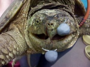
- Cut the excess wire gently using wire cutters and place a small epoxy ball over the exposed wire to prevent trauma to handlers.
- Flush and apply SSD to fracture lines and holes every other day for dry docked turtles or fractures that do not touch the water during a soak and every day for water turtles.
- Wire ties should stay in for 2-3 months until fractures completely stabilize and are healing well.
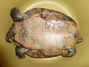
Note: Plastron fractures can be difficult to manage. Turtle Rescue Team has had some success by gluing paper clips to opposing edges of plastron fractures.
Relevant Videos
Other Surgical Procedures:
Additional Resources
Norton TM, Fleming GJ, and Meyer J. Shell surgery and repair. Chapter 113 in, Divers SJ and Stahl SJ (Eds.), Mader’s Reptile and Amphibian Medicine and Surgery, 3rd Ed., Amsterdam:Elsevier, 2019.
Divers SJ. Surgical equipment, instrumentation, and general principles. Chapter 90 in, Divers SJ and Stahl SJ (Eds.), Mader’s Reptile and Amphibian Medicine and Surgery, 3rd Ed., Amsterdam:Elsevier, 2019.
Kischinovsky M and Divers SJ. Ear. Chapter 92 in, Divers SJ and Stahl SJ (Eds.), Mader’s Reptile and Amphibian Medicine and Surgery, 3rd Ed. Amsterdam:Elsevier, 2019.
Key Concepts
- Turtle skin should be sutured closed using an interrupted horizontal mattress pattern. Muscle layers can be sutured using a simple continuous or interrupted pattern.
- Dremels, super glue, baking, soda, wire, bra hooks, and needle drivers can be used to set most carapace fractures.
- Aural abscesses fill with caseous pus and can be cleaned out to facilitate healing and make a turtle much more comfortable.
